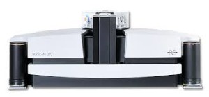
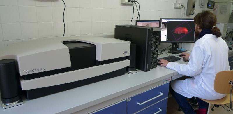
μCT Bruker SkyScan 1272
Technical features
X-ray source: 20-100 kV, 10 W
Detector: 16 Mp, cooled CCD fiber-optically coupled to scintillator
Resolution: <0.35 μm
Sample dimensions: max diameter: 75 mm; max length 70 mm
Heating and cooling stage from -30 to 85 °C
Material testing stage (compression and tension) of 42 and 440 N
Fast image reconstruction
Applications
High resolution μCT is very useful for the study of material structure, since it enables to have a 3D look (but also 2D) into the sample without breaking it. This technique has a wide range of applications, from medicine to material sciences, from soil science to biology and entomology, from archaeology to geology and does not require any pretreatment or preparation of the specimen. The instrument we have in the “Micro X-ray Lab” can also investigate the variations of the microstructure of samples when subjected to thermal and mechanical stresses, which is very important for material science studies.
